Glaucoma: Imaging Optic Nerve Damage
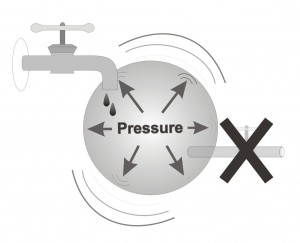
Glaucoma is a common disease, which usually occurs, in older age. Its incidence is 2-4% of the population older than 40, while in the 80+ year old population its incidence is greater than 6%. The optic nerve is damaged in glaucoma because of increased intra-ocular pressure, pressure that, over time, damages optic nerve fibers. Such damage to the nerve is irreversible. Glaucoma is the number two cause of blindness in Israel today. It is possible to treat glaucoma successfully, using medications in the form of eye drops, with laser surgery, and in very difficult cases, via surgery.
Most cases of blindness occur due to late diagnosis of the disease. In the majority of cases glaucoma occurs without signs of pain, and most patients do not sense a loss (narrowing) of their visual field. Therefore, unfortunately, a significant number of patients are diagnosed in the advanced stages of the disease, after many years of damage to the optic nerve due to untreated elevated intra-ocular pressure. There is a famous story of a well-known baseball player in the US who suffered unknowingly from advanced glaucoma. He was only diagnosed once he had lost all vision in one eye, while in the second eye he had significant damage to the nerve. The exposure of his story led to increased public awareness of the need for every one over forty to have an eye exam, even if the person has no symptoms or complaints. Screening tests are particularly important if any close family member suffers from glaucoma.
Recently several imaging instruments have been developed which allow us to measure the thickness of the retinal nerve fiber layer, which is the innermost of the ten retinal layers. This layer is composed of axons from ganglion cells, the same axons that unite to form the optic nerve. Because the thickness of this layer ranges from 30-150 microns, depending on the specific location in the retina, and because the layer is completely lucid, the ability to precisely measure this layer is a breakthrough in the diagnosis and monitoring of glaucoma patients.
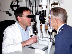
There are two methods to measure the severity of damage caused by glaucoma. Visual field examination gives an indication of functional damage, while the appearance of the optic nerve, and even more so, the retinal nerve fiber layer thickness, provide an indication of nerve tissue loss. While visual field is a subjective exam lasting 20-30 minutes, we can now image glaucomatous damage in an exam that lasts only a few minutes, does not require pupil dilation, and is completely objective.
At the Glaucoma Service, at Rambam’s Eye Department, chaired by Prof. Eytan Blumenthal, a new device has been incorporated, called a scanning laser polarimeter (GDx), which enables exact measurement of retinal fibers, aiding in the diagnosis of glaucoma. In addition, repeating measurements every few months to years enables us to monitor disease progression. This monitoring allows us to adjust treatment appropriately based on the severity of disease in each eye.
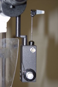
The instrument, called GDx (short for glaucoma diagnosis), scans the retina in approximately one second, creating a map detailing the retinal nerve fiber layer thickness of the central retina, in particular in the region surrounding the optic nerve. This imaging is based on a change in polarity of the light rays, polarity which changes as a function of retinal nerve fiber layer thickness in each and every spot on the retina. The machine provides, beyond actual thickness measurements, also a statistical analysis that indicates the level of damage in each area of the retina. This is the first GDx machine stationed in Israel, and it can enable improved diagnosis and monitoring of our glaucoma patients.

In figure 1. we see a series of GDx mapping, which simulates progressive damage from normality (left) to complete blindness (right).

Figure 2. is an example of mapping a glaucomatous eye.
A: Visual field which appears damaged in the lower region (pay attention that visual fields are “inverted”, such that damage in the lower half corresponds to damage to the upper half of the retina).
B: Optic nerve appearance as seen using the GDx, pay attention to the large cupping due to severe glaucoma.
C: GDx map showing retinal nerve fiber layer thickness at each retinal location.
D:Statistical analysis indicates severity of damage at each and every point. Pay attention to the fact that damage is also seen in the lower part, which the visual field test did not succeed in indicating (upper half of visual field looks normal in picture A).
E: Statistical analysis of the damage compared to normal values.
Literature:
- Blumenthal EZ and Weinreb RN. Assessment of the retinal nerve fiber layer. Surv Ophthalmol, 2001. 45: p. S305-12; discussion S332-4.
- Bowd C, Zangwill LM, Berry CC, Blumenthal EZ, Vasile C, Sanchez-Galeana C, Bosworth CF, Sample PA, Weinreb RN. Detecting early glaucoma by assessment of retinal nerve fiber layer thickness and visual function. Invest Ophthalmol Vis Sci, 2001. 42(9): p. 1993-2003.