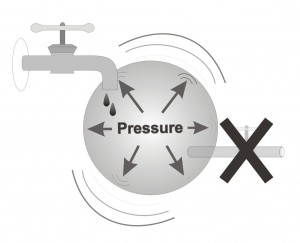
Glaucoma is a medical term referring to a group of eye diseases defined by elevated intraocular pressure which may result in irreversible damage to eye sight due to atrophy of the optic nerve.
The process of “seeing” begins when light rays enter the eye via the pupil and hit the retina, which is situated inside the globe, along the wall of the eye. Cells within the retina process this light into electrical signals using complicated bio-chemical reactions. These electrical signals are then transferred through nerve fibers which connect the eye to the brain. In the brain, yet another, complex and miraculous process occurs; the translation of the electrical signals into an image. This image is, in fact, what we see.
Each eye has one optic nerve, which connects it to the brain. It is through these nerves that information, which will eventually be interpreted into our vision, is passed to the brain. The ocular nerve is a few millimeters thick and a few centimeters long and it contains about a million nerve fibers. The optic nerve begins in a region called the optic disc. This area can be compared to the opening of a tube, which contains a large number of small ducts, each duct representing a single nerve fiber. The optic disc is the only part of the optic nerve, which can be directly observed by the ophthalmologist during an eye examination. By examining the optic disc, your ophthalmologist can determine if any injury or harm have occurred to the nerve fibers. Damage caused by Glaucoma is expressed as a loss (disappearing) of part of these nerve fibers.
In Glaucoma, damage (atrophy) occurs to ganglion cells, which are situated in the inner layer of the retina. The nerve fibers described earlier (which together make the optic nerve), are extensions of these ganglion cells. Loss of these cells (and their extensions) will, therefore, inevitably lead to atrophy (degeneration) of the optic nerve.
In Glaucoma, not all of the nerve cells die at once. Their loss (degradation) is slow and gradual, most often occurring over a period of many years. The typical order in which these cells atrophy and die is directly related to their place within the retina. Due to this cell loss the connection between the eye and the brain is slowly damaged. As fewer and fewer nerve fibers remain to transfer the information from the eye to the brain, the images formed in the brain are partial in comparison to the images formed in healthy people.
In conclusion an analogy can be made: the ocular nerve can be compared to an electrical cord containing many electrical wires (approximately a million). Each one of these fine wires begins within the retina and all end in the visual cortex of the brain. Each wire transports a small part of the picture to the brain, which then integrates this information together to produce a complete image. To those readers who are computers-competent, each fiber can be compared to one pixel in an image formed on a computer monitor screen (thus, each eye transmits to the brain a million pixels of visual information).
The next example will help to explain the damage caused to the vision by glaucoma. Imagine yourself looking at scenery. Now imagine looking at the same scenery through a hollow tube, 10cm in diameter. Even though the center of the picture remains identical, the circumference of the full picture will be missing. The circumference is called “the peripheral visual field”. As the diameter of the tube you are looking trough becomes narrower, your “central visual field” will become smaller. In glaucoma, usually the first ganglion cells to die are ones situated more peripheral in the retina. Most often, their absence is not noticeable, since the central visual field is not harmed and visual clarity is not affected. As the disease progresses, more peripheral ganglion cells die, causing the visual field to become narrower which results in “tunnel vision”, a syndrome which is typical of progressive glaucoma. This manifestation of the glaucomatous eye results from intact central visual field but a very impaired peripheral visual field. Most patients do not notice the loss of their peripheral visual field as long as their central visual field remains intact.
In other words, even with a loss of up to 90% or more of all retinal ganglion cells (and hence a similar loss of optic nerve fibers), the central visual field can remain intact, resulting in the disease going unnoticed. Therefore, even when the damage to the visual field is very advanced, the patient may not be aware of its presence at all. This is where the great danger of glaucoma lies; the patient may first notice problems with his vision only at a very advanced stage of the disease and if the disease is not treated early, there is dangers of further deterioration and even, in the worst of cases, complete irreversible blindness.
Your ophthalmologist can examine the retina and the optic disc as part of examining the back of the eye. When ganglion cells die as a result of glaucoma, their nerve fibers (also called axons) disappear. As a result the glaucomatous optic disc has a typical appearance. Glaucoma is therefore a disease in which irreversible damage results in the death of ganglion cells, which are required for transporting visual information from the eye to the brain.