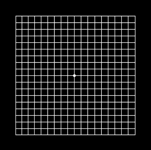
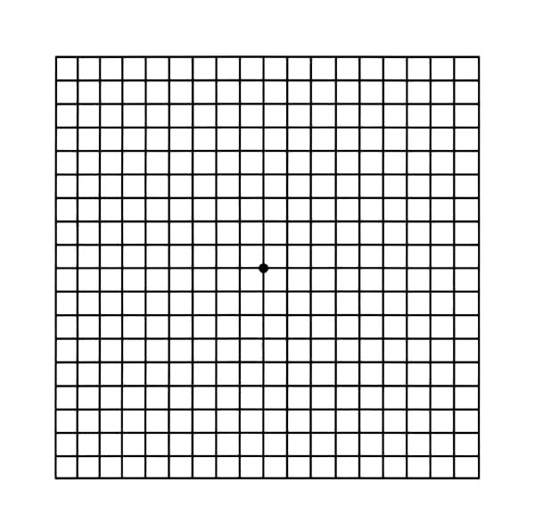
The Amsler grid is a quick and simple test that any patient can perform at home. Its purpose is to check the integrity of the central retina, known as the “macula”. The aim of this test is primarily to rule out the development of a disease known as Age-dependent Macular degeneration (AMD).
This very simple (and low-tech) diagnostic test allows the patient to ascertain whether the macula is functioning properly, or else, to determine the presence of a meaningful disturbance. This test may reveal the presence of any number of visual disturbances, including: distortion of the center of vision (macula and/or fovea), areas of deficient vision near the macula, and any number of conditions manifesting as distortion. The outcome of this test relates to the patient seeing straight lines as appearing crooked or bent, orderly squares appearing to be stretched out or distorted, or sections of lines appearing missing or discontinuous.
How to perform the Amsler Grid test
- If you use reading glasses (or bifocal/multifocal glasses which also serve as reading glasses), wear them.
- Look at the Amsler grid illustration (appearing on this computer screen) from a distance of approximately 40 cm (bent arm’s length, your habitual distance for watching the computer monitor).
- Cover one eye at a time, so that you are testing each eye separately (each time one eye will be checked while the other will be covered by the palm of your hand).
- Focus your attention on the central dot in the illustration and look at it for around 10 seconds.
How does one know if the test result is OK?
- If all the lines appear straight, and all the boxes seem equal in size, than the test went fine and there are no signs on the Amsler grid test to suggest macular damage.
- If, on the other hand, in a particular area of the picture, any of the lines appear crooked, broken, wavy, stop abruptly, or are blurry; or, if some of the similarly sized squares appear distorted (meaning that they are not shaped as squares, or that they appear to be stretched, distorted or differ in size from other squares), than the results of the Amsler grid examination exam indicates that damage to the macula might have occured.
***If the exam indicates damage for the first time, or if there is a change or worsening as compared to the previous time that you performed the test, it is highly advisable for you to see your ophthalmologist in order to ascertain, or rule out, any new or deteriorated condition involving the macular of that eye.***
Amsler grid:


Macular degeneration
The macula is a small, but extremely important, portion of the retina, also called the central part of the retina, which allows for sharp and clear vision of small details. While the entire retina can see, and hence we can see in several directions at once, it is the macula that enables us to see very clearly up front. Without the macular vision we would not have the ability to read, recognize faces, nor see objects in a clear, sharp way, both near and far. Damage to the macula is most often a result of aging and degeneration of this area. Loss of vision may assume either an acute, abrupt, or a gradual, slow, tempo. In some, very serious conditions, abnormal blood vessels may form. These blood vessels allow for leakage of fluid or blood under the macula and in these cases vision loss may occur rapidly.
Defective functioning of the macula will manifest as blurriness and/or distortion of central vision. Under these circumstances it is difficult or even impossible to read, sew, work on a computer or perform other tasks that require reasonable vision, such as recognizing faces. On the other hand, macular degeneration alone does not lead to full (total) blindness.
Symptoms that may be due to macular degeneration include:
- When reading a page, the word which one stares at (or a portion of that word), may appear blurry.
- Straight lines may appear crooked, interrupted, bent or curved, especially at the center of the field.
- Appearance of dark or blank areas in the center of vision.
Different optic tools can be used by individuals with impaired vision to allow them normal daily function:
- Magnification tools (magnifying glass, telescopes etc.)
- Closed-circuit television (which allows for great magnification of any image, such as text and photos).
- Special reading material printed in large type.
- Different computer and electronic appliances that are geared toward people with impaired vision.
Your ophthalmologist can recommend different devices or refer you to a specialist in vision rehabilitation. Since peripheral vision is not affected, this preserved vision may be the basis for visual rehabilitation towards the aim of reasonably normal daily function.
