A computerized visual field examination is a test that enables us to evaluate the amount of damage existing in the visual field of an eye suffering from glaucoma. If, on the other hand, no damage is found, we might conclude that the eye is either healthy, or at risk to develop future visual field damage.
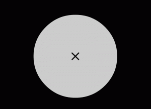
In this examination the patient’s field of vision is tested, while looking at a single fixed point in the center of the examination bowl. Visual field implies the ability to see the entire view in front of us. For example, while driving, we may look at the red light in front of us, but at the same time our peripheral (side) vision enables us to see a car pulling next to us in an adjacent lane.
Accurate testing of the visual field allows us to diagnose glaucoma at a very early stage, long before the patient actually notices any changes in his/her vision. Routine visual field examinations provide close monitoring of any progression in the disease.
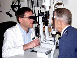
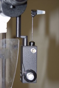
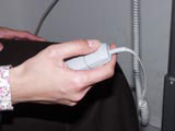
A computerized visual field examination is preformed as follows: the patient sits on a chair, and places his/her head on a chin-rest, and asked to look at a fixed light in front of him, without moving his/her eyesight from that little light situated in the middle of a large white bowl. One eye is covered with a patch, while the other eye takes the test. Afterwards, the patch is moved to the other eye, and the test is repeated for the remaining eye. While looking at the central light, additional faint white lights show up at different locations, each light presenting for a brief period of under a second. The patient is instructed to press a little button held in his/her hand each time he notices the faint white light. The faint light is projected at different intensities, some barely noticeable, and some not noticeable. No examinee can possibly recognize all the lights presented. The aim of the test is to identify at each visual field location what is the dimmest light that the person is able to see. All this information is later translated within the computer into a visual field map that shows the amount of glaucomatous damage existing in each eye.
The advantages of the computerized visual field test described above are that it is performed in a standardized manner each time, even if performed at the other end of the world. All these visual field devices perform equally, are calibrated the same way, and are barely influenced by the person delivering the test. This standardization enables your ophthalmologist, or glaucoma expert, to identify changes in your visual field, and learn from it whether your glaucoma is stable or progressing. That, in turn, will influence the treatment that you will receive, whether medical, laser, or surgical.
A basic explanation: What is a visual field test?
Examples of visual field test results:
| Normal visual field |  |
 |
| Glaucoma suspect |  |
 |
| Definite glaucoma but early |  |
 |
| Moderate damage |  |
 |
| Moderate damage |  |
 |
| Moderate-severe damage |  |
 |
| Moderate-severe damage |  |
 |
| Severe damage |  |
 |
| Very severe damage |  |
 |
| Very severe damage |  |
 |
| Near blindness |  |
 |